In the ever-advancing landscape of medical and scientific research, imaging techniques play a pivotal role in diagnosing diseases, understanding biological processes, and developing new treatments. The question of whether ligands can be used in imaging techniques is not only relevant but also holds significant promise for the future of these fields. As a trusted ligands supplier, we are at the forefront of providing high-quality ligands that can potentially revolutionize imaging methodologies.
Understanding Ligands
Before delving into their potential in imaging techniques, it's essential to understand what ligands are. Ligands are molecules that bind to specific receptors or molecules in the body. This binding is highly specific, much like a key fitting into a lock. The specificity of ligand binding is what makes them so valuable in biological and medical applications.
Ligands can be organic or inorganic molecules, and they come in a wide variety of shapes and sizes. Some common types of ligands include small molecules, peptides, proteins, and antibodies. Each type of ligand has its own unique properties and binding characteristics, which determine its suitability for different applications.
The Role of Ligands in Imaging
Imaging techniques rely on the ability to visualize specific structures or processes within the body. This can be achieved by using contrast agents or probes that interact with the target of interest. Ligands can serve as these contrast agents or probes by binding to specific receptors or molecules in the body, making them visible using various imaging modalities.
One of the most significant advantages of using ligands in imaging is their specificity. By targeting specific receptors or molecules, ligands can provide highly detailed and accurate information about the location and function of these targets. This can be particularly useful in diagnosing diseases, such as cancer, where specific receptors or molecules are often overexpressed or mutated.
Another advantage of using ligands in imaging is their ability to be labeled with imaging agents. For example, ligands can be labeled with radioactive isotopes, fluorescent dyes, or magnetic resonance imaging (MRI) contrast agents. These labels allow the ligands to be detected using different imaging modalities, such as positron emission tomography (PET), single-photon emission computed tomography (SPECT), fluorescence imaging, or MRI.
Types of Imaging Techniques Using Ligands
There are several types of imaging techniques that can utilize ligands. Here are some of the most common ones:
Positron Emission Tomography (PET)
PET is a powerful imaging technique that uses radioactive tracers to visualize metabolic processes in the body. Ligands labeled with positron-emitting isotopes, such as fluorine-18 or carbon-11, can be used as PET tracers. These tracers are injected into the body and then taken up by the target tissue or cells. The positrons emitted by the isotopes interact with electrons in the body, producing gamma rays that can be detected by a PET scanner. This allows for the visualization of the distribution and metabolism of the ligand in the body.
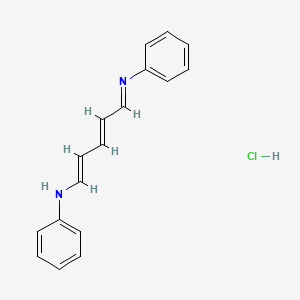
For example, Glutacondianil Hydrochloride丨CAS 1497-49-0 could potentially be labeled with a positron-emitting isotope and used as a PET tracer to target specific receptors or molecules in the body. This could provide valuable information about the function and distribution of these targets, which could be useful in diagnosing diseases or monitoring the effectiveness of treatments.
Single-Photon Emission Computed Tomography (SPECT)
SPECT is another imaging technique that uses radioactive tracers to visualize the function and structure of organs and tissues in the body. Ligands labeled with single-photon-emitting isotopes, such as technetium-99m or iodine-123, can be used as SPECT tracers. These tracers are injected into the body and then taken up by the target tissue or cells. The single photons emitted by the isotopes are detected by a SPECT scanner, which creates a three-dimensional image of the distribution of the tracer in the body.
Fluorescence Imaging
Fluorescence imaging is a non-invasive imaging technique that uses fluorescent dyes or proteins to visualize biological processes in the body. Ligands labeled with fluorescent dyes can be used as fluorescence probes. These probes are injected into the body and then taken up by the target tissue or cells. The fluorescent dyes emit light when excited by a specific wavelength of light, which can be detected by a fluorescence microscope or imaging system. This allows for the visualization of the distribution and function of the ligand in the body.
Magnetic Resonance Imaging (MRI)
MRI is a widely used imaging technique that uses magnetic fields and radio waves to create detailed images of the body. Ligands labeled with MRI contrast agents, such as gadolinium or iron oxide nanoparticles, can be used as MRI probes. These probes are injected into the body and then taken up by the target tissue or cells. The MRI contrast agents alter the magnetic properties of the surrounding tissue, which can be detected by an MRI scanner. This allows for the visualization of the distribution and function of the ligand in the body.
Challenges and Limitations
While the use of ligands in imaging techniques holds great promise, there are also several challenges and limitations that need to be addressed. One of the main challenges is the development of ligands with high specificity and affinity for their targets. This requires a deep understanding of the structure and function of the target receptors or molecules, as well as the ability to design and synthesize ligands that can bind to them effectively.
Another challenge is the development of imaging agents that can be safely and effectively labeled with ligands. This requires careful consideration of the properties of the imaging agent, such as its stability, toxicity, and pharmacokinetics. In addition, the labeling process needs to be optimized to ensure that the ligand retains its binding properties and specificity.
Finally, the cost and availability of ligands and imaging agents can also be a limitation. The development and production of high-quality ligands and imaging agents can be expensive, and they may not be readily available in all regions. This can limit the widespread use of ligands in imaging techniques, particularly in developing countries.

Our Role as a Ligands Supplier
As a ligands supplier, we are committed to providing high-quality ligands that can be used in imaging techniques. We offer a wide range of ligands, including Glutacondianil Hydrochloride丨CAS 1497-49-0, Diphenyl-2-pyridylphosphine丨CAS 37943-90-1, and Trihexylamine丨CAS 102-86-3, which can be used for a variety of applications, including imaging.
Our ligands are synthesized using state-of-the-art techniques and are rigorously tested to ensure their purity, quality, and stability. We also offer custom synthesis services to meet the specific needs of our customers. In addition, we work closely with our customers to provide technical support and guidance on the use of our ligands in imaging techniques.
Conclusion
In conclusion, ligands have the potential to play a significant role in imaging techniques. Their specificity and ability to be labeled with imaging agents make them valuable tools for visualizing specific structures and processes within the body. While there are still challenges and limitations that need to be addressed, the future of using ligands in imaging looks promising.
As a ligands supplier, we are excited to be part of this growing field and are committed to providing high-quality ligands that can help advance the development of imaging techniques. If you are interested in learning more about our ligands or would like to discuss potential applications in imaging, please feel free to contact us. We look forward to working with you to explore the possibilities of using ligands in imaging and to contribute to the advancement of medical and scientific research.
References
- Weissleder, R., & Pittet, M. J. (2008). Imaging in the era of molecular oncology. Nature, 452(7187), 580-589.
- Blasberg, R. G., & Patlak, C. S. (1997). Principles of tracer kinetic analysis for the study of blood-brain barrier function and substrate transport. In Cerebral blood flow and metabolism (pp. 133-162). Lippincott-Raven.
- Licha, K., & Kiessling, F. (2005). Molecular imaging: from bench to bedside. European Journal of Radiology, 56(2), 167-174.